The bony structure of the pelvis creates the foundation for the hip joint. The pelvis is formed of the fusion of the lateral ilium, lateral ischium, and the anterior pubis. To the posterior, the pelvis articulates with the sacrum. As an intact complex, the pelvis looks like an inward-sloping structure. The word “pelvis” thus comes from the Latin word for basin.
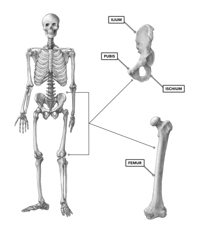
Figure 1: The bones of the hip and pelvis
Each side of the pelvis has a hip joint anchored to the vertebral column by way of the sacroiliac joint between the ilium and sacrum.
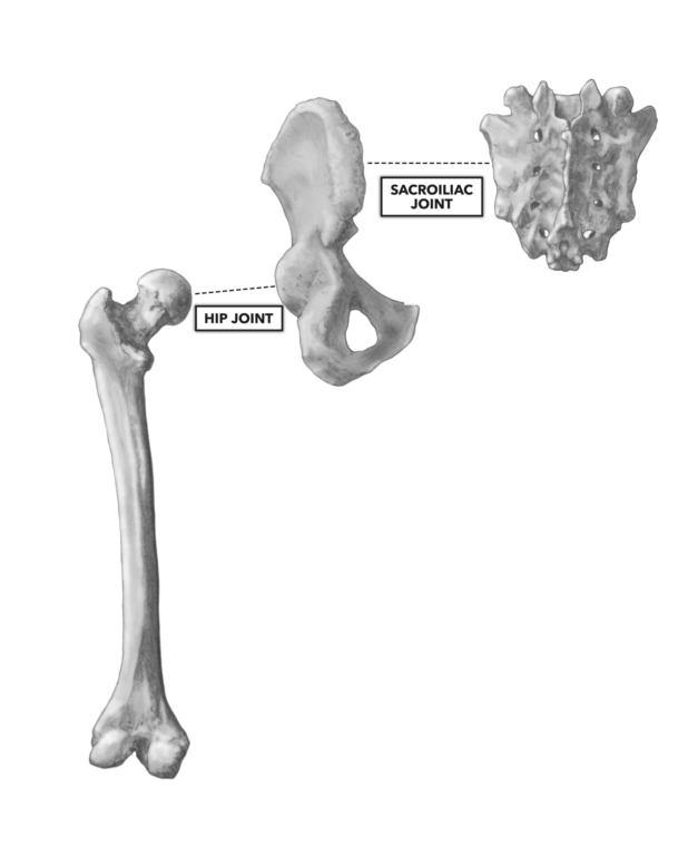
Figure 2: The sacroiliac joint
In general, we think of the hip joint as the place where the femur of the thigh articulates with the pelvis, one bone connected to the other. However, the hip joint is the articulation of four bones: (1) the femur, (2) pubis, (3) ilium, and (4) ischium. The latter three are connected by the triradial cartilage in a manner very similar to the suture joints of the skull.
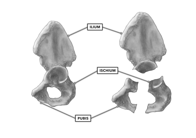
Figure 3: The ilium and ischium
The hip is a synovial, multi-axial ball-and-socket joint, the largest of its type in the human body. Its diameter in adults is approximately two inches. Formed by the articulation of the head of the femur and a relatively deep fossa in the pelvis known as the acetabulum, the hip joint has a very mobile but fairly stable structure. Due to its heavy musculature and robust architecture, hip dislocations in life and sport are fairly rare.
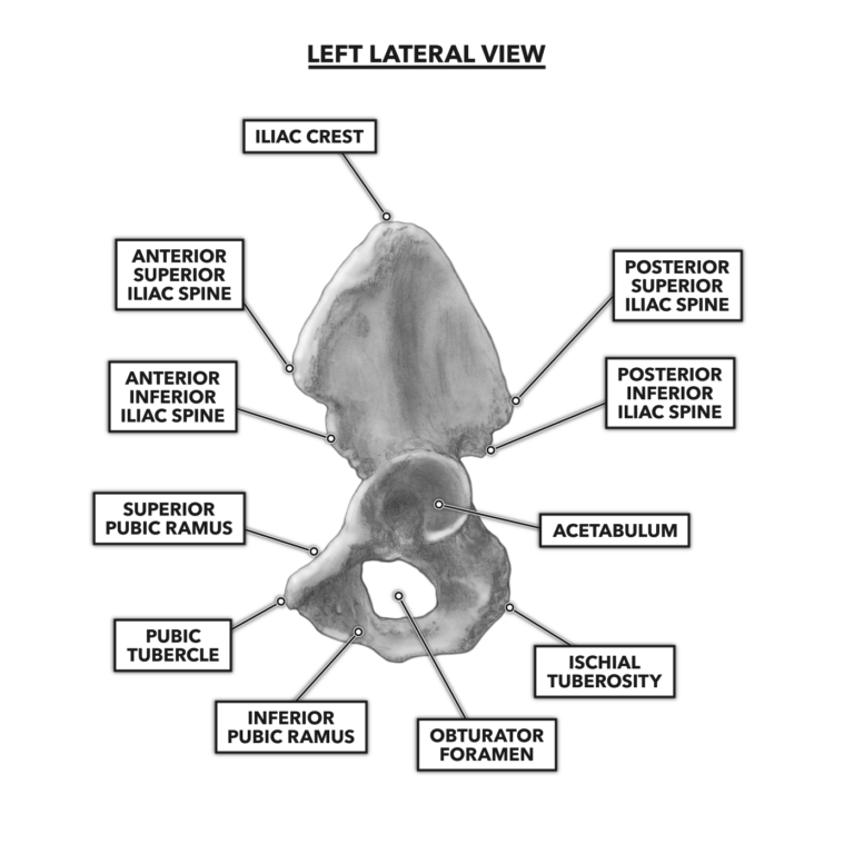
Figure 4: Lateral view of the hip
The pelvis provides many attachment points for muscles and ligaments. The pelvis structure also acts to protect soft tissues (nerves, blood vessels, urinary tract, reproductive organs, etc.).
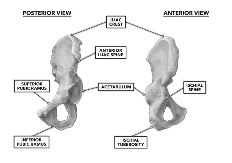
Figure 5: Anterior and posterior views of the hip
The single most important aspect of the hip joint’s structure is its articulation with the proximal femur, where it acts as a transition and connector between the vertebral column and legs. The architecture of this joint enables the upper body to be supported while sitting, standing, and during ambulation.
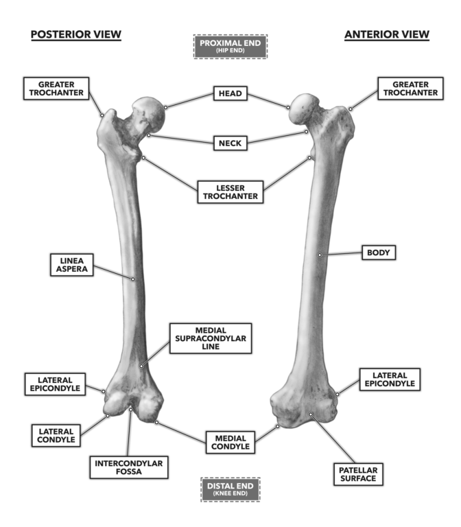
Figure 6: Anterior and posterior views of the femur
To learn more about human movement and the CrossFit methodology, visit CrossFit Training.
Bones of the Hip & Pelvis